Toni Susin
Last modified 03/04/2013
Menu:
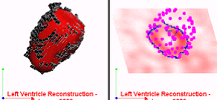
LEFT VENTRICLE RECONSTRUCTION FROM SPECT IMAGES.
Researches: Oscar García Panyella, Antonio Susin
 Description:
Our approach describes the three-dimensional reconstruction of the internal and external surfaces of the human’s left ventricle from actual SPECT data. The reconstruction is a first process fitting in a complete VR application that will serve as an important diagnosis tool for hospitals. Beginning with the surfaces reconstruction, the application will provide volume and interactive real-time manipulation with the model. We focus on speed, precision and smoothness for the final surfaces. As long as heart diseases diagnosis requires experience, time and professional knowledge, simulation is a key-process that enlarges efficiency.
Access to a 3D model obtained from patient’s data can have several applications like support on diagnosis, surgery planning, student’s training or even remote-operation. A first approximation to the problem would be using a manual process with specific image-processing software though it would require deep medical knowledge and experience. Efficiency can be improved via simulation. In this sense we present a dynamic model designed to fit regions formed from soft tissue. We use the evolution of a deformable mesh affected by internal and external forces. Internal forces are defined in terms of elasticity; external forces are derived from the images data set as a vector field called GVF. The evolution from an initial elliptic mesh under these forces has been studied with different models of elasticity ranging from free particles, mass-spring and plane elasticity. Different numerical integration methods have been tested.

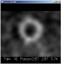
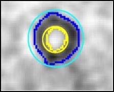
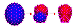
Fig 2. Reconstruction process
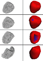
We have several recoveries with non-missing data and partial missing data, performed using different fillings for a PHANTOM test volume. We have made some recovering test experiments with 10%, 32% and 53% percentages of missing volume data, always referred to the 100% of the total. The best recovery is the second one because it gives a percentage of missing volume of 29.1% against the 32% of the real data emptied. This represents a relative percentage error of 0.09. For the other test examples we obtain relative errors of 0.45 and 0.24 respectively.
 |
Volume=265501 mm3 |
Volume=250944 mm3 |
|
Volume=188360 mm3 |
|
Volume=159970 mm3 |
Fig. 3. Percentage of volume recovered
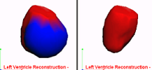
We also present a reconstructed cardiac cycle from an actual patient data, all along with its associated ejection fraction estimation. Ejection fraction value is estimated using medical software and compared with our system evaluation.

Fig. 4. Reconstructed cardiac cycle
 The reconstruction of both the internal and external surfaces, allowed us to present the synthetic cardiac cycle for an actual patient and derive its associated ejection fraction estimation.
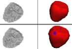
The segmentation module has been improved in order to provide a better and robust border detection and classification. Now the filtering process is based on the Maximum-Likelihood Classification approach (MLC), which rewards an edge depending on its probability to be a part of the left ventricle. The algorithm also takes into account vertical coherence between slices that improves the inputs adding information on the 3D shape.
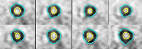
In terms of practical results, we test all these new features against several left-ventricle reconstructions applied to actual pathological hearts.
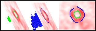
Figure 5. The segmented left ventricle. From left to right: internal (3D),
external (3D) and both borders (2D).

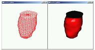
Fig. 6 Complete pipeline of the geometry reconstruction from the Spect images.
García O., Susín A. Two Strategies on Border Labeling applied to the Estimation of Ejection Fraction. 4es. Jornades de Recerca en Enginyeria Biomèdica. (2004) Barcelona (Spain).(PDF 363Kb)
García O., Susín A. MLC filtering applied to the 3D reconstruction of the left ventricle.
Proc. CEIG 2003, XIII Congreso Español de Informática Gráfica. pp 1–12 (2003) (PDF 810Kb)
García O., Susín A. Left Ventricle Volume Estimation From 3D SPECT Reconstruction.
IEEE Computers in Cardiology 2002. Ed. A. Murray A vol. 29, pp 621–624 (2002) (PDF 500Kb)
García O., Susín A. Surface and Volume Reconstruction of the Left Ventricle from SPECT data.
in procc. 1st Iberoamerican Symposium in Computer Graphics (SIACG 2002)July 2-5, 2002 Guimarães, Portugal (PDF 682Kb)

García O., Susín A. Left Ventricle's Surface Reconstruction and Volume Estimation.
3es. Jornades de Recerca en Enginyeria Biomèdica. Lacroix D., Ginebra M.P. ed pp.1--10 (2002)
Xarxa Temàtica en Enginyeria Biomèdica (ISBN 84-699-8705-4). Vic (Spain) (PDF 270Kb)
García O., Susin A.Modelo Dinámico para la reconstrucción del Corazón Humano
XVII CEDYA, VII Congreso de Matemática Aplicada. L.Ferragut, A. Santos ed. pp 733-734 (2001) Univ. Salamanca, Spain (spanish PDF 129Kb)
García O., Susin A., Navazo I. Segmentación automática mediante un modelo dinámico. Aplicación a la reconstrucción del ventrículo izquierdo 2ones. Jornades de Recerca en Enginyeria Biomèdica. Giraldo B., Caminal P., ed. pp.173--176 (2000)
Xarxa Temàtica en Enginyeria Biomèdica (ISBN 84-607-1365-2). Sitges (Spain) (PDF 169Kb)
Oscar García Pañella.
Deformable Models for Left Ventricle Reconstruction using Spect Images.
PhD dissertation: Dept. de Tecnología y Multimedia. Univ. Ramón Llull 24/03/2004.
References:
[Bro-Nielsen94] Morten Bro-Nielsen: “Active Nets and Cubes”, IMM Tech. Rep 94-13, 1994.
[Canny86] J. Canny. “A Computational Approach to Edge Detection”, IEEE Transactions on Pattern Analysis and Machine Intelligence, Vol. 8, No. 6, Nov. 1986.
[Cohen93] L. D. Cohen and I. Cohen, “Finite-Element Methods for Active Contour Models and Balloons for 2-D and 3-D”, IEEE Transactions. PAMI, vol. 15, no. 11, pag. 1131-1147, 1993.
[Kass88] M. Kass, A. Witkin, and D. Terzopoulos, “Snakes: Active Contour Models”, International Journal of Computer Vision, vol. 1, no. 4, pag. 321-331, 1988.
[Lorensen87] W. Lorensen and H. Cline, “Marching Cubes: A High Resolution 3D Surface Construction Algorithm”, Proceedings. SIGGRAPH’87, pag. 163-169, 1987.
[Mannting95] Finn Mannting, Puneet K Chandak, Yanina V Zabrodina, B Leonard Holman, “Atlas of Myocardial Perfusion SPECT”, Brigham and Women’s Hospital, Harvard Medical School, Boston MA. 1995-1997.
[McInerney94] T. McInerney and D. Terzopoulos, “A Dynamic Finite Element Surface Model for Segmentation and Tracking in Multidimensional Medical Images with Application to Cardiac 4D Image Analysis”, Journal of Computerized Medical Imaging and Graphics, 1994.
[Montagnat00] Johan Montagnat, Hervé Delingette, Nicolas Scapel i Nicholas Ayache, “Representation, shape, topology and evolution of deformable surfaces. Application to 3D medical image segmentation”, Rapport de Recherche, Mai 2000, INRIA.
[Park96] Jinah Park, “Model-Based shape and motion analysis: left ventricle of a heart”, A dissertation in Computer and Information Science. University of Pennsylvania, 1996.
[Quackenbush96] D. Quackenbush, P. Ratiu, and J. Kerr, “Segmentation of the Visible Human Data Set”, The Visible Human Project Conference, pag. 69-70, 1996.
[Sakaue96] K. Sakaue, “Stereo Matching by the Combination of Genetic Algorithm and Active Net”, Systems and Computers in Japan, vol. 27, no. 1, pag. 40-48, 1996.
[Takanashi98] I. Takanashi, S. Muraki, A. Doi, and A. Kaufman, “3D Active Net for Volume Extraction”, Proc. SPIE Vol. 3298, p. 184-193, Visual Data Exploration and Analysis V, Robert F. Erbacher; Alex Pang; Eds. 5/1998
[Xu97] C. Xu and J.L. Prince, “Gradient Vector Flow: A New External Force for Snakes”, IEEE Proc. CVPR, 1997.
[Xu98] C. Xu and J.L. Prince, “Snakes, Shapes, and Gradient Vector Flow”, IEEE Transactions on Image Processing, pag. 359-369, 1998.